ShopDreamUp AI ArtDreamUp
Deviation Actions


Description
Thanks everyone for your kind comments  (Smile)") This
This  was featured in DA front page and it received many comments I need sometime to reply all the comments so in the meanwhile, enjoy this photo
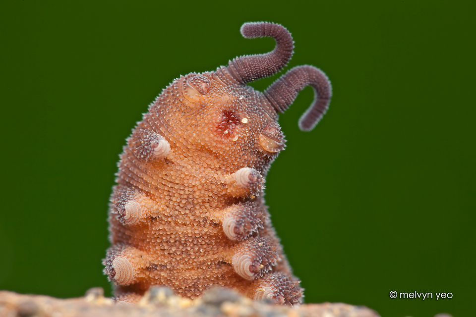
Taken at night in Singapore forest.
Watch how it hunts!!! www.youtube.com/watch?v=FbVDYS…
Quote from en.wikipedia.org/wiki/Onychoph…
The stub feet that characterise the velvet worms are conical, baggy appendages of the body, which are internally hollow and have no joints. Although the number of feet can vary considerably between species, their structure is basically very similar. Rigidity is provided by the hydrostatic pressure of their fluid contents, and movement is usually obtained passively by stretching and contraction of the animal's entire body. However, each leg can also be shortened and bent by internal muscles. Due to the lack of joints, this bending can take place at any point along the sides of the leg.
In some species, two different organs are found within the feet:
- Crural glands are situated at the shoulder of the legs, extending into the body cavity. They open outwards at the crural papillae—small wart-like bumps on the belly side of the leg—and secrete chemical messenger materials called pheromones. Their name comes from the Latin cruralis meaning "of the legs".
- Coxal vesicles are pouches located on the belly side of the leg, which can be everted and probably serve in water absorption. They are only found within the family Peripatidae and are named from coxa, the Latin word for "hip".
On each foot is a pair of retractable, hardened (sclerotised) chitin claws, which give the taxon its scientific name: Onychophora is derived from the Greek onyches, "claws"; and pherein, "to carry". At the base of the claws are three to six spiny "cushions" on which the leg sits in its resting position and on which the animal walks over smooth substrates. The claws are used mainly to gain a firm foothold on uneven terrain.
Apart from the pairs of legs, there are three further body appendages, which are at the head and comprise three segments:
Antennae - On the first head segment is a pair of slender antennae, which serve in sensory perception. They probably do not correspond directly to the antennae of the Arthropoda, but perhaps rather with their "lips" or labrum. At their base is found a pair of simple eyes, except in a few blind species. In front of these, in many Australian species, are various dimples, the function of which is not yet clear. It appears that in at least some species, these serve in the transfer of sperm-cell packages (spermatophores).
Mouthparts - On the belly side of the second head segment is the labrum, a mouth opening surrounded by sensitive "lips". In the velvet worms, this structure is a muscular outgrowth of the throat, so, despite its name, it is probably not homologous to the labrum of the Arthropoda. Deep within the oral cavity lie the sharp, crescent-shaped "jaws", or mandibles, which are strongly hardened and resemble the claws of the feet, with which they are probably homologous;[6]:146 early in development, the jaw appendages have a similar position and shape to the subsequent legs. The jaws are divided into internal and external mandibles and their concave surface bears fine denticles. They move backward and forward in a longitudinal direction, tearing apart the prey.
The surface of the mandibles is smooth, with no ornamentation.[8] The cuticle in the mandibles (and claws) is distinct from the rest of the body. It has an inner and outer component; the outer component has just two layers (whereas body cuticle has four), and these outer layers (in particular the inner epicuticle) are dehydrated and strongly tanned, affording toughness.
Slime glands - On the third head segment, to the left and right of the mouth, are two openings designated "oral papillae". Within these are a pair of large, heavily internally branched slime glands. These lie roughly in the centre of the body and secrete a sort of milky-white slime, which is used to ensnare prey and for defensive purposes. Sometimes the connecting "slime conductor" is broadened into a reservoir, which can buffer pre-produced slime. The slime glands themselves are probably modified crural glands.
All three structures correspond to an evolutionary origin in the leg pairs of the other segments.
was featured in DA front page and it received many comments Taken at night in Singapore forest.
Watch how it hunts!!! www.youtube.com/watch?v=FbVDYS…
Quote from en.wikipedia.org/wiki/Onychoph…
The stub feet that characterise the velvet worms are conical, baggy appendages of the body, which are internally hollow and have no joints. Although the number of feet can vary considerably between species, their structure is basically very similar. Rigidity is provided by the hydrostatic pressure of their fluid contents, and movement is usually obtained passively by stretching and contraction of the animal's entire body. However, each leg can also be shortened and bent by internal muscles. Due to the lack of joints, this bending can take place at any point along the sides of the leg.
In some species, two different organs are found within the feet:
- Crural glands are situated at the shoulder of the legs, extending into the body cavity. They open outwards at the crural papillae—small wart-like bumps on the belly side of the leg—and secrete chemical messenger materials called pheromones. Their name comes from the Latin cruralis meaning "of the legs".
- Coxal vesicles are pouches located on the belly side of the leg, which can be everted and probably serve in water absorption. They are only found within the family Peripatidae and are named from coxa, the Latin word for "hip".
On each foot is a pair of retractable, hardened (sclerotised) chitin claws, which give the taxon its scientific name: Onychophora is derived from the Greek onyches, "claws"; and pherein, "to carry". At the base of the claws are three to six spiny "cushions" on which the leg sits in its resting position and on which the animal walks over smooth substrates. The claws are used mainly to gain a firm foothold on uneven terrain.
Apart from the pairs of legs, there are three further body appendages, which are at the head and comprise three segments:
Antennae - On the first head segment is a pair of slender antennae, which serve in sensory perception. They probably do not correspond directly to the antennae of the Arthropoda, but perhaps rather with their "lips" or labrum. At their base is found a pair of simple eyes, except in a few blind species. In front of these, in many Australian species, are various dimples, the function of which is not yet clear. It appears that in at least some species, these serve in the transfer of sperm-cell packages (spermatophores).
Mouthparts - On the belly side of the second head segment is the labrum, a mouth opening surrounded by sensitive "lips". In the velvet worms, this structure is a muscular outgrowth of the throat, so, despite its name, it is probably not homologous to the labrum of the Arthropoda. Deep within the oral cavity lie the sharp, crescent-shaped "jaws", or mandibles, which are strongly hardened and resemble the claws of the feet, with which they are probably homologous;[6]:146 early in development, the jaw appendages have a similar position and shape to the subsequent legs. The jaws are divided into internal and external mandibles and their concave surface bears fine denticles. They move backward and forward in a longitudinal direction, tearing apart the prey.
The surface of the mandibles is smooth, with no ornamentation.[8] The cuticle in the mandibles (and claws) is distinct from the rest of the body. It has an inner and outer component; the outer component has just two layers (whereas body cuticle has four), and these outer layers (in particular the inner epicuticle) are dehydrated and strongly tanned, affording toughness.
Slime glands - On the third head segment, to the left and right of the mouth, are two openings designated "oral papillae". Within these are a pair of large, heavily internally branched slime glands. These lie roughly in the centre of the body and secrete a sort of milky-white slime, which is used to ensnare prey and for defensive purposes. Sometimes the connecting "slime conductor" is broadened into a reservoir, which can buffer pre-produced slime. The slime glands themselves are probably modified crural glands.
All three structures correspond to an evolutionary origin in the leg pairs of the other segments.
Image size
960x640px 377.5 KB
© 2013 - 2024 melvynyeo
Comments231
Join the community to add your comment. Already a deviant? Log In
WHAT ARE YOU?!